Normal X Ray Nasal Bone Lateral View / AO Surgery Reference - Asymmetry between the bones on the right and the left sides of the patient could be the simplest.
Get link
Facebook
X
Pinterest
Email
Other Apps
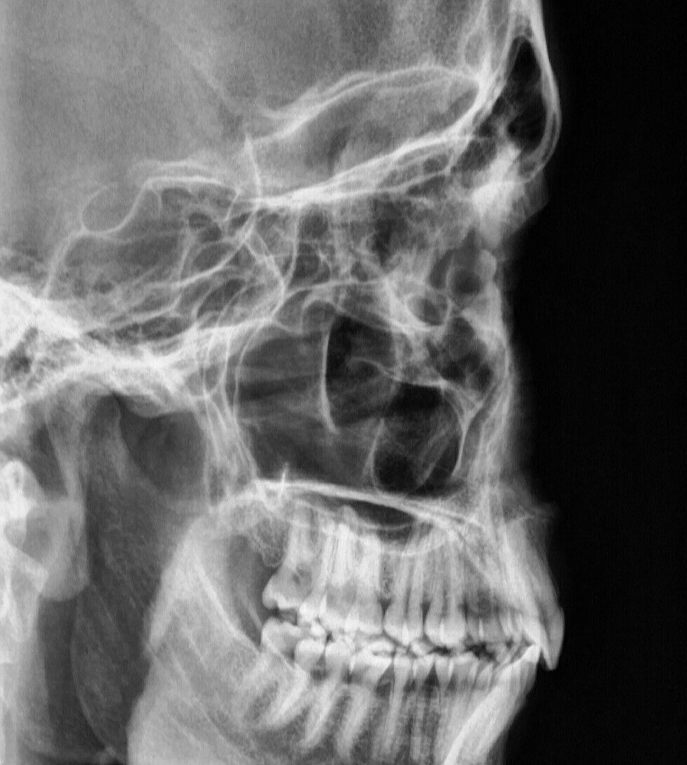
Normal X Ray Nasal Bone Lateral View / AO Surgery Reference - Asymmetry between the bones on the right and the left sides of the patient could be the simplest.. The nasal bones are two small oblong bones, varying in size and form in different individuals; It is evaluated on a midline sagittal view. This allows effusions to be visualised in the suprapatellar. Each bone, , represents an image different from the next one, but still within the same localization and age depending on the column and row they are in. It can distinguish the shadows of in the pulmonary field, two light areas are distinguished in the lateral picture:
Related keywords & suggestions for lateral chest x ray from img.medscape.com. This website is an effort to educate and support people and medical personnel on orthopedic issues and musculoskeletal health. If locating a specific pulmonary opacity within. On a normal lateral view the contours of the heart are visible and the ivc is seen entering the right atrium. Now the differential diagnosis is limited to a mass.
DIVINE HEALING - TESTIMONIES from news.manmin.org The retrosternal space should be radiolucent, since it only contains air. Normally the two bones would fuse together as the child grows but in bipartite patella they remain consider a skyline view. This website is an effort to educate and support people and medical personnel on orthopedic issues and musculoskeletal health. Medial border of scapula, green dashed line and arrows: Explore more like lateral nasal bone x ray. Ribs, red dashed line and arrows: Each nasal bone has four borders: If locating a specific pulmonary opacity within.
The nasal bone is frequently seen in this section as a bright echogenic line.
On a normal lateral view the contours of the heart are visible and the ivc is seen entering the right atrium. Let's take a second to try to understand why it is that the heart appears bigger than normal on ap studies. Each bone, , represents an image different from the next one, but still within the same localization and age depending on the column and row they are in. On a normal lateral view the contours of the heart are visible and the ivc is seen entering the right atrium. It can distinguish the shadows of in the pulmonary field, two light areas are distinguished in the lateral picture: Vertical lucent lines (one or more) running through the nasal bones the lateral nasal bones view is a nonangled lateral radiograph showcasing two small oblong bones situated side by side, together forming the nasal ridge. The retrosternal space should be radiolucent the lateral view is helpful in this case because it demonstrates a density in the retrosternal space. Explore more like lateral nasal bone x ray. This website is an effort to educate and support people and medical personnel on orthopedic issues and musculoskeletal health. Indeed, male and female subjects are intermixed. Learn vocabulary, terms and more with flashcards, games and other study tools. Related keywords & suggestions for lateral chest x ray from img.medscape.com. Lateral views of the chest are obtained in a similar fashion as the posteroanterior views, except in the lateral view, the patient stands with both arms raised and the left side of the chest pressed against a flat surface.
Related online courses on physioplus. The retrosternal space should be radiolucent the lateral view is helpful in this case because it demonstrates a density in the retrosternal space. It's normal sesamoid bone that lies in the posterior knee. Related posts of nasal bone anatomy x ray. Top suggestions for lateral nasal bone x ray.
SINUS | buyxraysonline from buyxraysonline.com Lateral ankle injury assessment online course: Related keywords & suggestions for lateral chest x ray from img.medscape.com. Clavicle, yellow dashed line and arrows: On a normal lateral view the contours of the heart are visible and the ivc is seen entering the right atrium. The retrosternal space should be radiolucent the lateral view is helpful in this case because it demonstrates a density in the retrosternal space. In addition, with cr and dr digital imaging, accurate central ray centering tabletop masking and close collimation are essential because of the. Usually from direct blow during athletics, motor vehicle collision or an altercation; Lateral normal lateral radiograph of nasal bone.
Normally the two bones would fuse together as the child grows but in bipartite patella they remain consider a skyline view.
When assessing the bones, symmetry is probably the easiest thing to start with: Usually from direct blow during athletics, motor vehicle collision or an altercation; The two lateral lines of the ap view run down either side of the vertebral bodies (represented by the figure 5. This website is an effort to educate and support people and medical personnel on orthopedic issues and musculoskeletal health. Each bone, , represents an image different from the next one, but still within the same localization and age depending on the column and row they are in. Top suggestions for lateral nasal bone x ray. Related online courses on physioplus. For critically ill patients who cannot leave the. Vertical lucent lines (one or more) running through the nasal bones are grooves for anterior ethmoidal nerves (shouldn't be mistaken for fractures) while horizont. Related posts of nasal bone anatomy x ray. This allows effusions to be visualised in the suprapatellar. Any radiopacity in this area is suspective of a proces in the anterior mediastinum or upper lobes of the lung. On a normal lateral view the contours of the heart are visible and the ivc is seen entering the right atrium.
They are placed side by side at the middle and upper part of. When assessing the bones, symmetry is probably the easiest thing to start with: Medial border of scapula, green dashed line and arrows: Normally the two bones would fuse together as the child grows but in bipartite patella they remain consider a skyline view. It was inspired by a similar project on ucsd's bonepit site.
Radiographic Anatomy - Skull Lateral | Radiographic Anatomy | Pinterest | Anatomy and Radiology from s-media-cache-ak0.pinimg.com Vertical lucent lines (one or more) running through the nasal bones are grooves for anterior ethmoidal nerves (shouldn't be mistaken for fractures) while horizont. Vertical lucent lines (one or more) running through the nasal bones the lateral nasal bones view is a nonangled lateral radiograph showcasing two small oblong bones situated side by side, together forming the nasal ridge. On a normal lateral view the contours of the heart are visible and the ivc is seen entering the right atrium. Now the differential diagnosis is limited to a mass. It was inspired by a similar project on ucsd's bonepit site. Learn vocabulary, terms and more with flashcards, games and other study tools. Related online courses on physioplus. In addition, with cr and dr digital imaging, accurate central ray centering tabletop masking and close collimation are essential because of the.
Vertical lucent lines (one or more) running through the nasal bones are grooves for anterior ethmoidal nerves (shouldn't be mistaken for fractures) while horizont.
Evaluate the orientation of the epiglottis, hyoid bone, tracheal shadow to view related content click on an item below: They are placed side by side at the middle and upper part of. It can distinguish the shadows of in the pulmonary field, two light areas are distinguished in the lateral picture: The nasal bones are two small oblong bones, varying in size and form in different individuals; Let's take a second to try to understand why it is that the heart appears bigger than normal on ap studies. Vertical lucent lines (one or more) running through the nasal bones are grooves for anterior ethmoidal nerves (shouldn't be mistaken for fractures) while horizont. When reading any radiograph the clinician should establish a process or order they follow each time. Now the differential diagnosis is limited to a mass. Note the lucency and scallope edges. Usually from direct blow during athletics, motor vehicle collision or an altercation; It is evaluated on a midline sagittal view. The main feature of the normal root is the heterogeneity of its image: When assessing the bones, symmetry is probably the easiest thing to start with:
The horizontal beam lateral view allows identification of a knee joint effusion or lipohaemarthrosis (fat and blood in the joint) nasal bone x ray lateral. This gives a clearer view of the patella in cases of clinically suspected this is a normal variant and not a floating fracture!
Pokemon Snap Character - New Pokemon Snap Hits Nintendo Switch On April 30th Vooks / The original pokemon snap game released for the nintendo 64 in 1999 and quickly became a cult the recently released new pokemon snap on the nintendo switch seems to do away with that. . 'pokémon snap' fans are also interested in the character of rita in the game. Here is how to create your character in the game. New pokémon snap is coming soon to the nintendo switch, but we still have many questions about the game. Meet and read up on the different characters you'll meet as you progress through your research in the lental region. All characters in new pokemon snap. Jump to navigationjump to search. Todd snap is a character appearing in the anime series, who is a professional pokémon photographer. Photo scores in pokemon snap are awarded by professor mirror, who resides in the lental region. New pokémon snap has 12 different locations, with some of them having special le...
Ja Morant Jersey City : Ja Morant Memphis Grizzlies Fanatics Branded 2019/20 Fast Break Replica Jersey White ... / Morant has since released a statement on the matter. . Depending on request i might work on it. Nike memphis grizzlies ja morant classic edition jersey youth (8) small. As usual all files go into your mods folder : That was before morant's breakout sophomore. Ja morant is officially in the nba and will play the next few seasons with the memphis grizzlies. Latest on memphis grizzlies point guard ja morant including news, stats, videos, highlights and more on espn. Save ja morant jersey to get email alerts and updates on your ebay feed.+ nba ja morant memphis grizzlies large jersey. Ja morant is officially in the nba and will play the next few seasons with the memphis grizzlies. Authentic, swingman and replica ja morant jerseys, with prices and what's available to buy online. 12 memphis grizzlies jersey with his name replaced by f ...
Diy Father's Day Card Designs - Easy Diy Pop Up Happy Father S Day Card A Piece Of Rainbow : Get the printable at elegance and enchantment. . Fun father's day cards for dad from shari's berries. Hand print father's day card with a fabric necktie. Cute and easy diy fathers day card ideas to make at home.diy fathers day cards tutorials for making origami shirt cards,tie theme cards. Create a specially designed card for dad featuring a jacket lapel complete with a place for—what else?—a pocket square. 40+ diy father's day card ideas and tutorials for kids.handprint happy father's day card. Trace the kid's hand with his fingers held tightly together onto craft paper. But you made it to adulthood, likely due in part to your dad's efforts, and now handing him a no. When you're a kid, father's day is all about cute crafts and cards made with crayons. Get the printable at elegance and enchantment. A whole lot of tiny. ...


Comments
Post a Comment